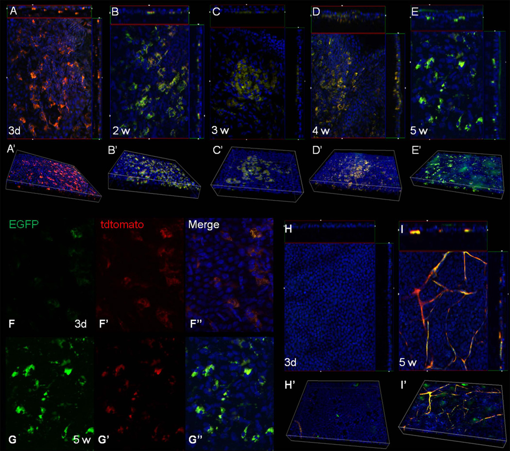
Figure 4.
Induction of EGFP expression from the HFSC transplant. Corneas were harvested at various times following transplantation from mice fed doxycycline chow and examined for the expression of red and green fluorescent protein via whole mount imaging. A Z-stack through the entire thickness was obtained for each sample, with each slice having a thickness of 1.8 µM. (A–E) Images represent the cut-view of the Z-stack depicting the x-z axis along the top and the y-z axis along the right-hand side of the x-y image. A 3-D reconstruction of the Z-stack is shown below each cut-view image (A’–E’). Three days following transplant most of the HF bulge-derived SC expressed the red fluorescent protein (mT) indicating they had not yet differentiated (A). The presence of EGFP was observed two weeks post-transplantation (B) and was maintained throughout the length of the experiment; 3 wk (C), 4 wk (D), 5 wk (E). At all time points, EGFP was localized to the basal layer of the corneal epithelium. (F–G) Single color images taken from the Z-stack depicting the cellular localization of mT and EGFP at 3 d (F) and 5 wk (G). (H, I) Cut-view images and the corresponding 3-D images of corneas from mice not receiving the HFSC transplant 3 d (H, H’) and 5 w (I, I’) following LSCD. Magnification: 200X

