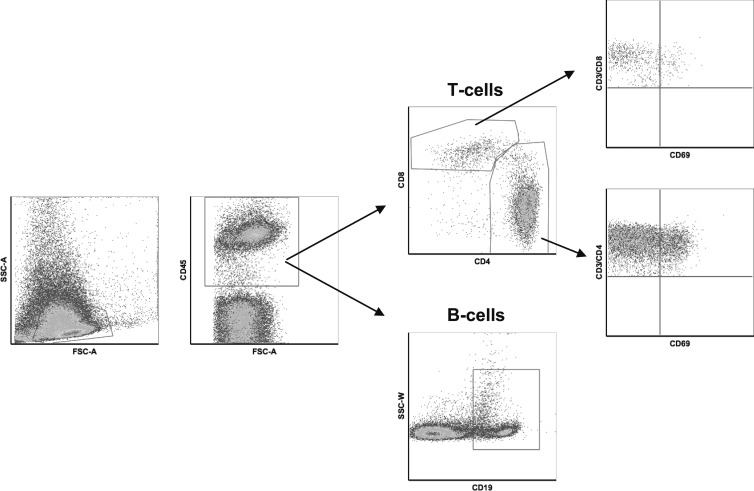
Figure 1.
Flow cytometry gating strategy. Fresh lymph node biopsies were immediately processed for flow cytometry analysis using T-cell surface markers including CD3, CD4, CD8, CD45 and CD69, and B-cell surface marker CD19. This figure shows the regions used to identify the different cell populations.

