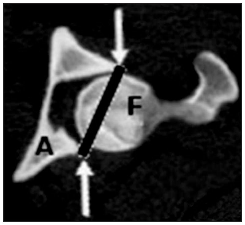
Figure 2.

Two-dimensional CT image showing identification of the surface plane of the acetabulum (A) in canine hip joints. Lines from the ventral to the dorsal aspect of each acetabulum (arrows) extending across the femoral head (F) on each slice permitted generation of a plane over the lateral surface of the acetabulum in the 3-D model.
