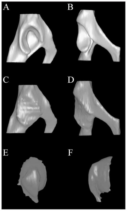
Figure 3.
Lateral (A) and ventrodorsal (B) views of a 3-D CT model of a canine acetabulum. The acetabular cavity was filled to the level of the surface plane as shown in lateral (C) and ventrodorsal (D) views. A so-called 3-D cast of the acetabulum was created by subtracting the bony acetabulum from the filled joint space as shown in medial (E) and dorsoventral (F) views. The casts have been rotated approximately 180° from their position in the acetabulum.

