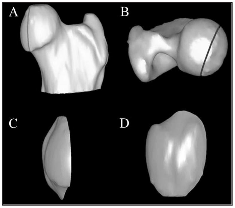
Figure 6.

Craniocaudal (A) and dorsoventral (B) views of a 3-D CT model showing measurement of the FVIA in a canine hip joint by use of data collected in Figure 5. The reference line from Figure 5 was used to isolate the portion of the femoral head within the acetabular cavity on the 3-D image. Craniocaudal (C) and mediolateral (D) views of the same model have been rotated approximately 45° and 90°, respectively, from their position relative to the proximal aspect of the femur.
