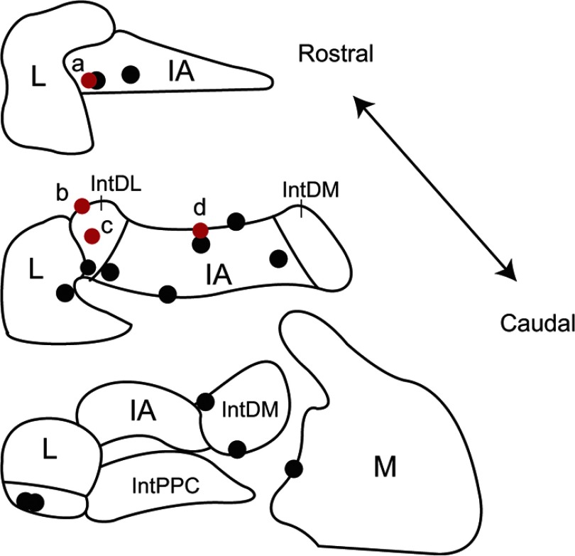
Figure 9.
Locations of recorded CN neurons. Locations of CN neurons with (red circles) and without (black circles) synaptic input from recorded crus 2a PCs as identified by analysis of CS–CN correlograms. Schematics of coronal sections of CN based on the atlas of Paxinos and Watson (1998) are shown. IA, Interpositus anterior; IntDL, dorsolateral division of interpositus; IntDM, dorsomedial division of interpositus; IntPPC, posterior parvicellular division of interpositus; L, lateral nucleus; M, medial nucleus.

