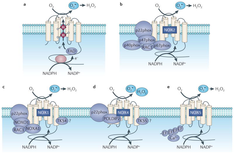
Figure 1. Structure and molecular organization of the NADPH oxidases of the NOX family.
Part a shows the topology and the enzymatic reaction catalysed by the NADPH oxidase (NOX) enzymes. Parts b–e represent the molecular structure of NOX2, NOX1, NOX4 and NOX5, which are predominantly expressed in carcinoma cells. All NOX proteins can form a complex with p22phox, but the cytosolic subunits differ between the NOX oxidase isoforms. e, electron; FAD, flavin, superoxide; POLDIP2, DNA polymerase-δ-interacting adenine dinucleotide; H, Haem; H2O2, hydrogen peroxide; O2 protein 2; TKS, tyrosine kinase substrate.

