Figure 3. Axonal loss and basal lamina redundancy in skin biopsies from the patient with PMP22 null.
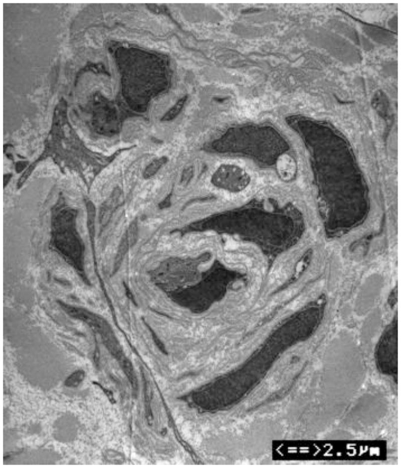
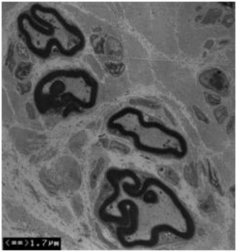
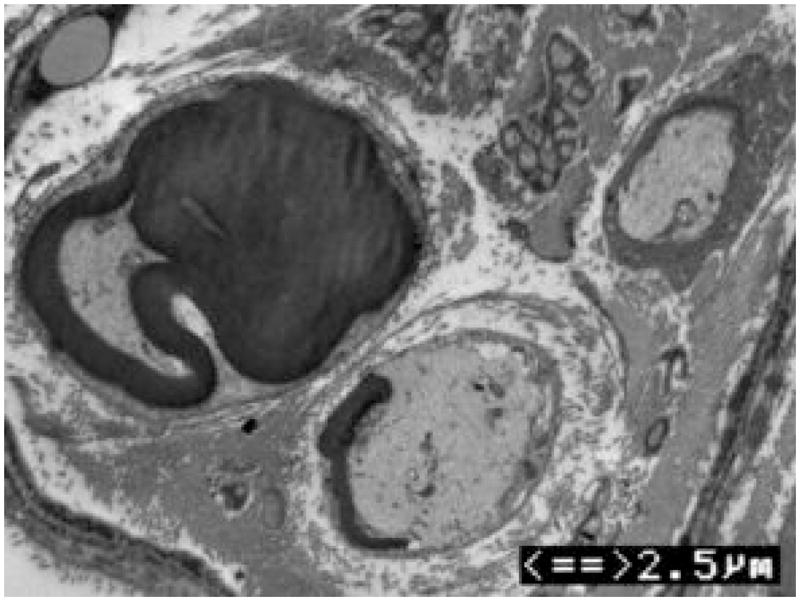
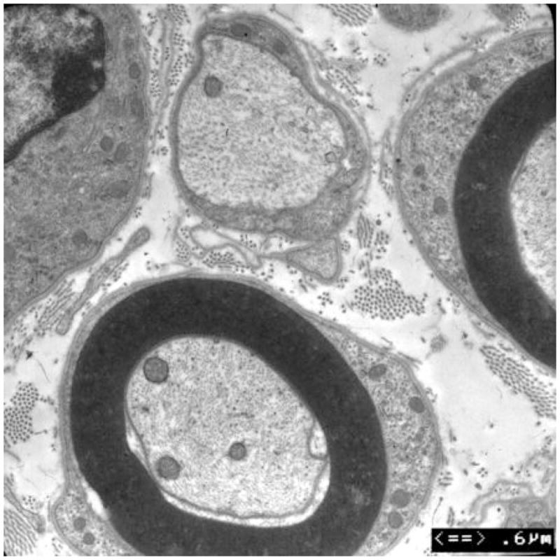
(A) Electron microscopy of the patient’s skin biopsy reveals Schwann cell processes loosely wrapping around an area where the axon appears to have degenerated. The overall appearance resembles an onion bulb. (B) The area circled by asterisks in A is enlarged to reveal redundant basal lamina of the Schwann cells (arrows). (C) A representative skin biopsy from a healthy control demonstrates the compacted myelin with normal thickness. (D) Skin biopsy of a pmp22−/− mouse reveals tomaculous formation (asterisk). Tomacula could not be identified in the patient’s dermal nerves since myelinated nerve fibers are degenerated. Two axons on the right side fail to form compact myelin. E. EM of pmp22−/− mouse sciatic nerve reveals a number of relatively large axons devoid of compact myelin, as exemplified by the axon on the top. In contrast, an axon in the bottom has a comparable diameter to the one on the top but its myelin is well formed. The excessive basal membrane redundancy is readily identifiable (arrow).
