
Figure 10.
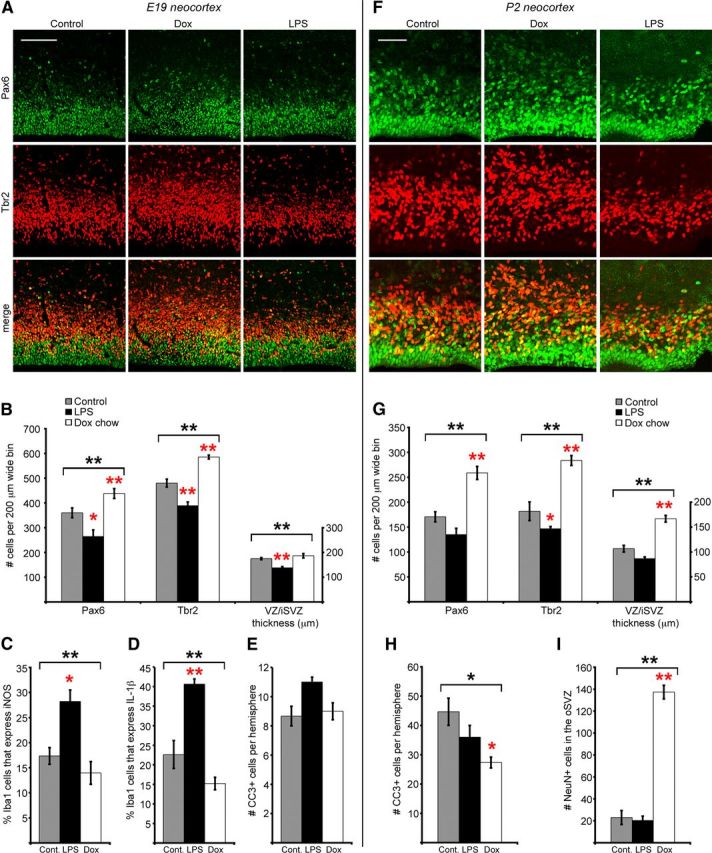
Manipulating the activation state of microglia changes the number of neural precursor cells in the prenatal brain. The number of Pax6+ and Tbr2+ precursor cells was quantified in 200-μm-wide bins that spanned from the ventricular surface through the proliferative zones at E19 and at P2. A, B, At E19 Dox treatment significantly elevated the number of Tbr2+ cells per bin, while LPS treatment significantly reduced the number of Pax6+ and Tbr2+ cells per bin, and reduced the combined thickness of the VZ/iSVZ. C, D, LPS treatment increased the proportion of Iba1+ cells that express high levels of iNOS, a marker of activated microglia (Verney et al., 2010), and the proportion of Iba1+ cells that expressed high levels of the cytokine IL-1β. The proportion of Iba1+ cells that expressed these markers was slightly decreased in the Dox group. E, LPS slightly elevated the number of CC3+ cells per hemisphere, but the difference was not significant. F, G, At P2 Dox treatment significantly increased the number of Pax6+ and Tbr2+ cells and increased the combined thickness of the VZ/iSVZ, while LPS significantly decreased the number of Tbr2+ cells. H, Dox treatment significantly reduced the number of CC3+ cells per hemisphere. I, Dox treatment significantly increased the number of NeuN+ cells in the oSVZ. Brackets denote ANOVA significance, *p < 0.05; **p < 0.01. Red asterisks indicate post hoc significance, single red asterisk: p < 0.05; double red asterisk: p < 0.01. Error bars show SE. Scale bars: A, 100 μm; B, 50 μm.