
Figure 3.
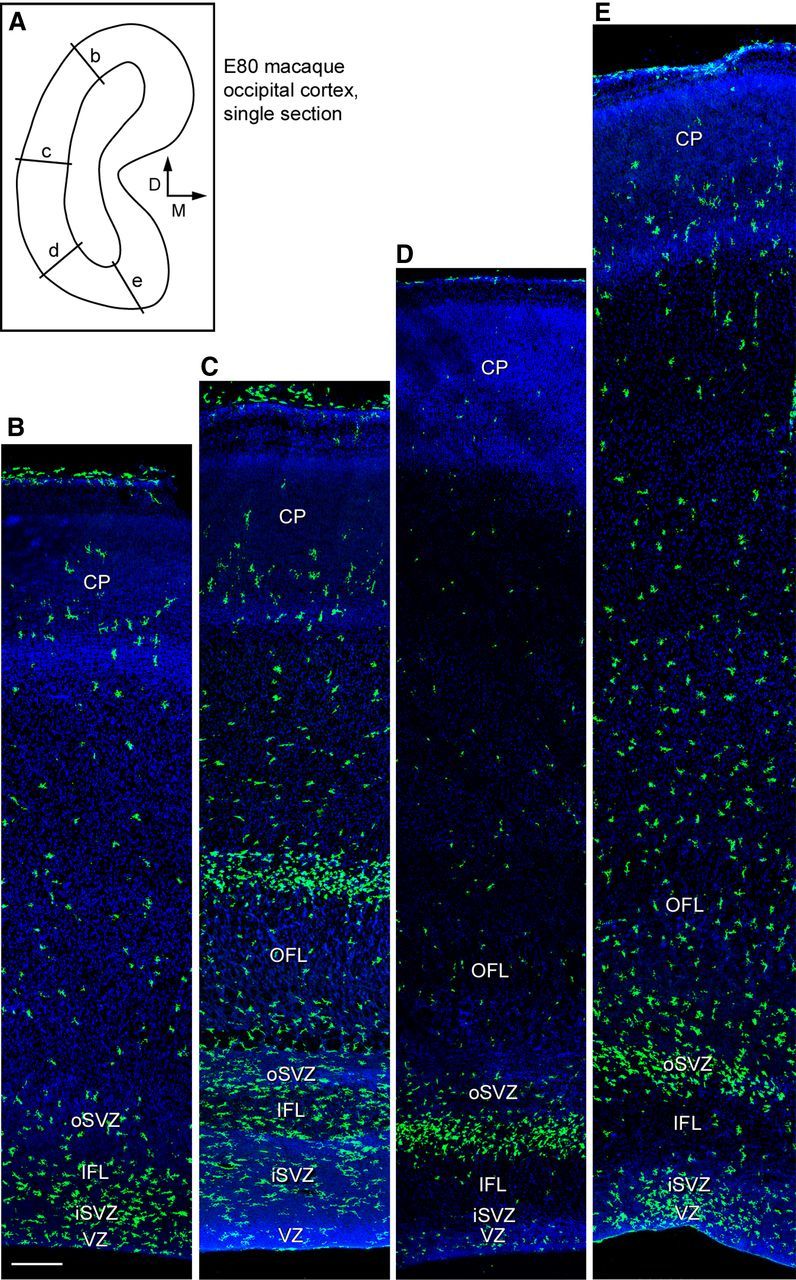
Iba1+ microglia distribution varies across cortical areas. A, Line drawing of a single coronal section of E80 macaque occipital cortex that was stained for Iba1 and confocal imaged to produce the images shown in B–E. Location of panels indicated by labeled lines, and orientation is indicated with arrows. B–E, Iba1+ microglia (green) colonize the iSVZ and oSVZ in the occipital cortex, but the position of the dense Iba1+ cell bands varies across cortical areas. Scale bar: (in B) B–E, 250 μm. Blue, DAPI.