
Figure 1.
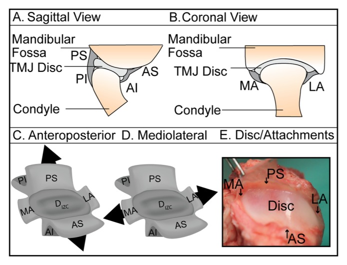
Schematic of TMJ discal attachments. (A) Sagittal and (B) coronal views of the attachments, including the medial (MA) and lateral (LA) attachments, and the posterior and anterior attachments, which bifurcate to form the posterior superior (PS), posterior inferior (PI), anterior superior (AS), and anterior inferior (AI) attachments. Each attachment region was tested in the (C) anteroposterior and (D) mediolateral directions. (E) Excised disc/attachment complex imaged prior to the isolation of specimens for testing.