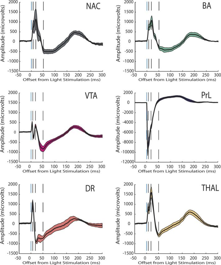
Figure 9.

Evoked potential recorded from limbic brain areas during low-frequency cortical stimulation (0.1 Hz). Evoked potentials are shown as the mean ± SEM for all of the LFP channels recorded across each structure within a single mouse during a single trial. This mouse had recording electrodes implanted in dorsal raphe (DR) (AP, −4.5 mm; ML, 0.3 mm; and DV, −2.25 mm from bregma) and medial dorsal thalamus (THAL) (AP, −1.6 mm; ML, 0.3 mm; and DV, −2.9 mm from bregma) in addition to the other brain areas described in this study. Note that several brain areas that were most distal to the stimulation site (i.e., DR and VTA) exhibited maximum/minimum potential peaks that occurred before the proximal brain areas (i.e., NAc).
