Figure 3.
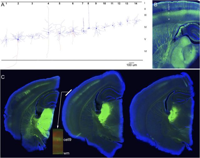
Histological verification of recorded cells and ChR2 expression. A, Identification of some of the cells studied. In some cases, during histological processing, the apical dendrite was truncated and lost by the resectioning (80 μm) of the 400 μm slices. B, Typical thalamocortical slice obtained from a Thy1-ChR2-eYFP-line18 mouse (1 resectioned 80 μm section is shown). EYFP, green; DAPI, blue. Note the robust eYFP fluorescence (ChR2 expression) of layer V cells and their apical dendrites, and the typical sparseness of expression in layer IV, indicating lack of ChR2 expression in thalamocortical fibers. C, Three successive slices from a CD-1 mouse cut in the thalamocortical plane (1 resectioned 80 μm section is shown) that had been injected with AAV-hSyn-ChR2-eYFP in somatosensory thalamus. Note the robust ChR2 expression of thalamocortical fibers in layer IV, and somewhat in layer VI, of somatosensory cortex. The inset strip in the middle slice shows a neurobiotin-filled cell (red) recorded in that slice overlapped with the ChR2 expression (green); this is cell9 reconstructed in A. EYFP, green; DAPI, blue; DyLight594, red. wm, white matter.

