


Figure 1.
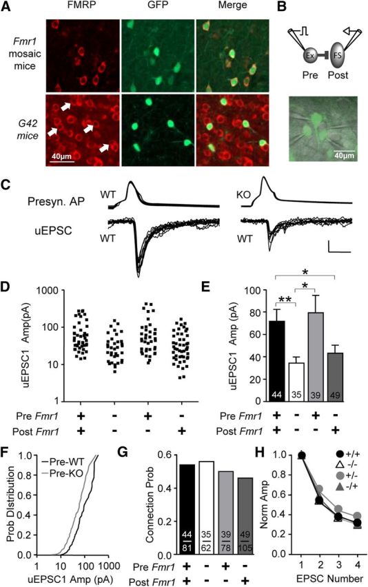
Presynaptic deletion of Fmr1 in mosaic mice results in decreased unitary excitation of FS inhibitory neurons. A, Immunohistochemistry for GFP and FMRP in somatosensory neocortex at P14. Top, In GFP/Fmr1 mosaic mice, GFP marks neurons that express FMRP, while lack of GFP indicates no FMRP. Bottom, In WT mice expressing GFP in parvalbumin-positive FS neurons, FMRP colocalizes with GFP indicating that FS neurons express FMRP (arrows). B, Top, Diagram of glutamatergic connection examined. Bottom, In live mosaic slices, GFP-positive and GFP-negative neurons were clearly identified. C, Example traces obtained from simultaneous recordings show that uEPSCs observed in FS neurons and evoked from presynaptic KO excitatory neurons were smaller. Calibration: 50 mV, 20 pA; 5 ms. D, E, Scatter plot and bar graph showing that average uEPSC1 amplitude was smaller whenever Fmr1 was deleted from the presynaptic excitatory neuron. F, Cumulative distribution of uEPSC1 amplitude for merged presynaptic KO and merged presynaptic WT groups. G, No detectable change in connection probability was observed. H, Average normalized uEPSC amplitude during the train of four presynaptic action potentials was unchanged. *p < 0.05; **p < 0.01.