


Figure 9.
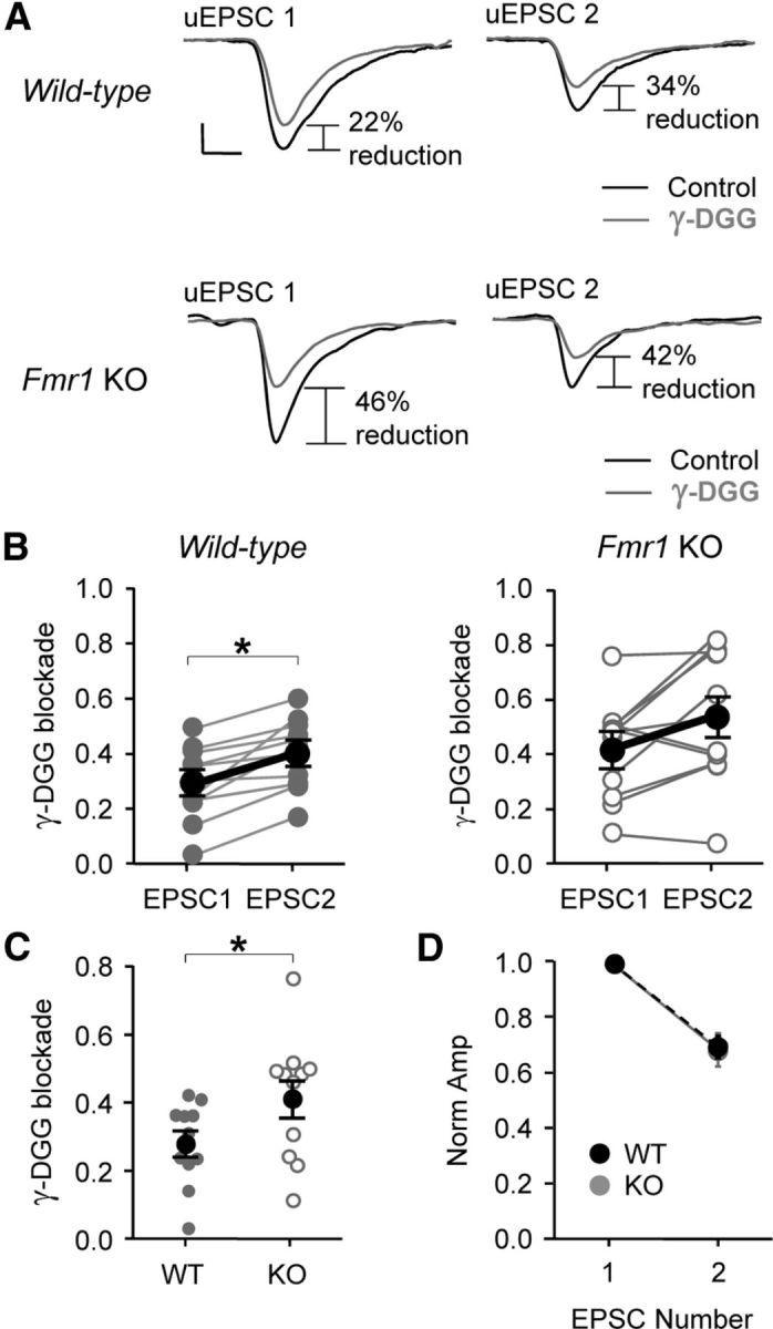
Decreased multivesicular release exists in the constitutive Fmr1 KO. A, Example average traces from Fmr1 WT and KO slices demonstrating the blockade caused by the AMPAR competitive antagonist, γ-DGG (500 μm). Note the greater blockade at uEPSC2 when compared to blockade at uEPSC1, indicating multivesicular release. B, Paired line plots showing blockade for uEPSC1 and uEPSC2 in Fmr1 WT (left) and KO slices (right). Dark lines indicate averaged data. Calibrations: 20 pA, 2 ms. C, Average blockade for uEPSC1 was greater in KO slices indicating reduced multivesicular release. D, No difference observed in short-term plasticity. *p < 0.05.