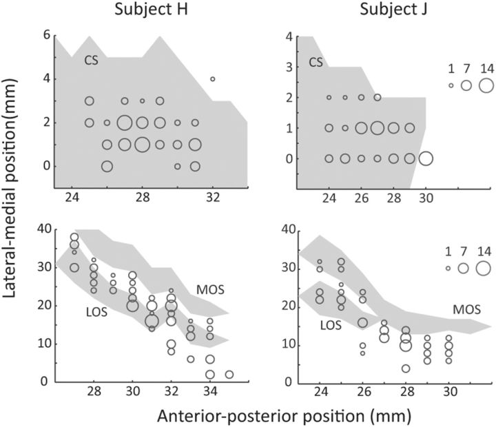
Figure 2.
Number of neurons recorded at each location in ACC (top) and OFC (bottom). The anterior–posterior position is measured from the interaural line. In subject H, the genu of the corpus callosum was at AP 24 mm and in subject J it was at AP 23 mm. In the ACC plot, the lateral–medial position extended from the fundus of the cingulate sulcus (0 mm) to more medial positions within the dorsal bank of the cingulate sulcus. In the OFC plot, the lateral–medial position extended from the ventral bank of the principal sulcus (0 mm), around the inferior convexity, and onto the orbitofrontal surface. The extent of sulci is shown by the gray shading. CS, Cingulate sulcus; MOS, medial orbital sulcus; LOS, lateral orbital sulcus.

