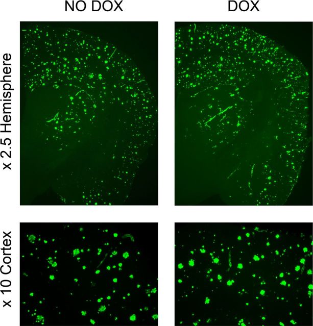
Figure 4.
Persistence of thioflavin-positive amyloid plaques in APPsi:tTA mice. After the completion of all behavioral tasks, the mice were intracardially perfused with cold PBS under anesthesia, and half-brains were fixed by immersion in 4% paraformaldehyde. The 30 μm frozen coronal sections through the cortex and hippocampus were stained with the amyloid-binding dye Thioflavin S. The images shown are representative of the entire cohort of animals that were behaviorally tested.

