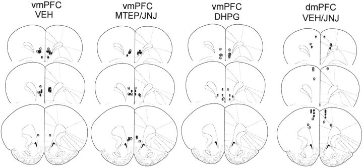
Figure 6.
Histological verification of microinjector placements within the PFC. A diagram depicting the results of our histological examination of microinjector placements conducted on the completion of testing for the residual effects of an intra-vmPFC infusion of VEH, MTEP, JNJ 16259685 (JNJ), and DHPG and of an intra-dmPFC infusion of JNJ 16259685, as examples of placements for the animals included in the statistical analyses of the data in this study. As exemplified by the saline (open circles) and cocaine (filled circles) animals in this figure, only animals exhibiting microinjector placement within the ventral prelimbic cortex, within the infralimbic cortex or at their interface, were included in the statistical analyses of the data for vmPFC studies. As exemplified by the VEH (open circles) and JNJ 16259685 (filled circles) animals in the panel for the dmPFC study, only animals exhibiting microinjector placement within the dorsal prelimbic cortex, the anterior cingulate, or their interface were included in the statistical analyses of the data for the dmPFC study.

