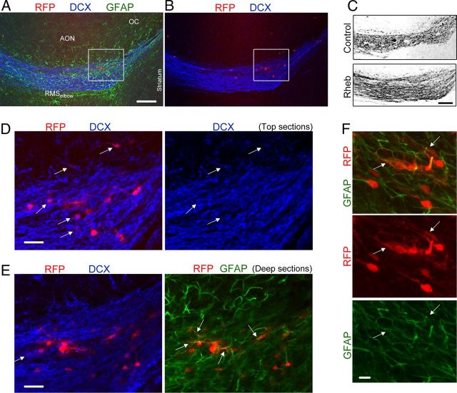
Figure 3.
Heterotopia contained neuroblasts, astrocytes, and cells with a neuronal morphology at P19. A, B, Coimmunostaining for GFAP (green) and DCX (blue) in a sagittal section containing RhebCA-expressing cells at the RMSelbow. C, Confocal image of DCX immunostaining in the RhebCA and control conditions. Image for the RhebCA condition is the same as that shown in A and B. D, E, Immunostaining for GFAP (green) and DCX (blue) of the cells shown in the white square in A and B. The top and deep sections are shown in D and E, respectively. The arrows point to RFP+ cells being DCX+ or GFAP+. F, Immunostaining for GFAP (green) of RhebCA-expressing cells in a heterotopia at the RMSelbow. The arrows point to RFP+ cells being GFAP+. Scale bars: A–C, 150 μm; E, 30 μm; F, 10 μm.

