
Figure 5.
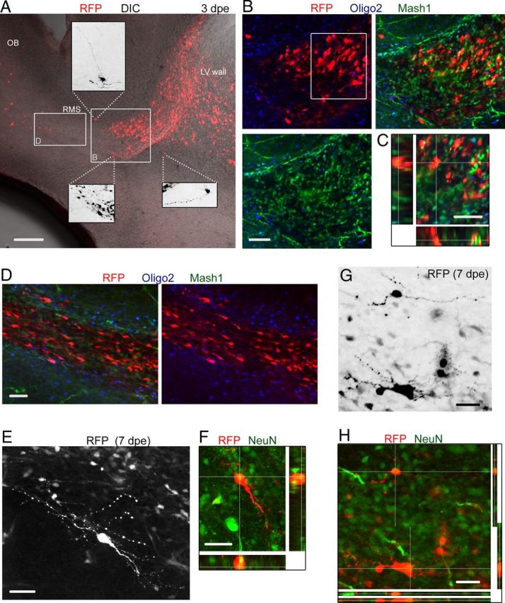
RFP+ cells at early neonatal stages include Mash1+ cells, neuroblasts, and differentiating neurons in the RhebCA condition. A, Confocal image of a sagittal slice at 3 dpe (P4) containing RFP+ cells in the RhebCA condition. Cells are already seen entering the OB circuitry. Insets, Zooms of some ectopic RFP+ cells with a more differentiated morphology. DIC, Differential interference contrast; LV, lateral ventricle. B, Coimmunostaining for Olig2 (blue) and Mash1 (green) of RFP+ cells shown in the white square in A. C, One Z section and multiple Z-associated projections for RFP+ cells that are Mash1+ from the white square in B. D, Confocal image of Mash1 and Olig2 immunostaining in the RMS from the white rectangle shown in A. Neuroblasts are Olig2− and Mash1−. E, F, Confocal images of a differentiated cell at 7 dpe (P8) that stained for NeuN (green) as shown in the projections in F. E shows a Z-stack, and F shows a single Z. G, Confocal image of RFP+ cells in the RMSelbow. H, Immunostaining for NeuN (green) showing that RFP+ cells with a more differentiated morphology are NeuN+. Scale bars: A, 150 μm; B, G, H, 50 μm; C, 40 μm; D–F, 70 μm.