
Figure 1.
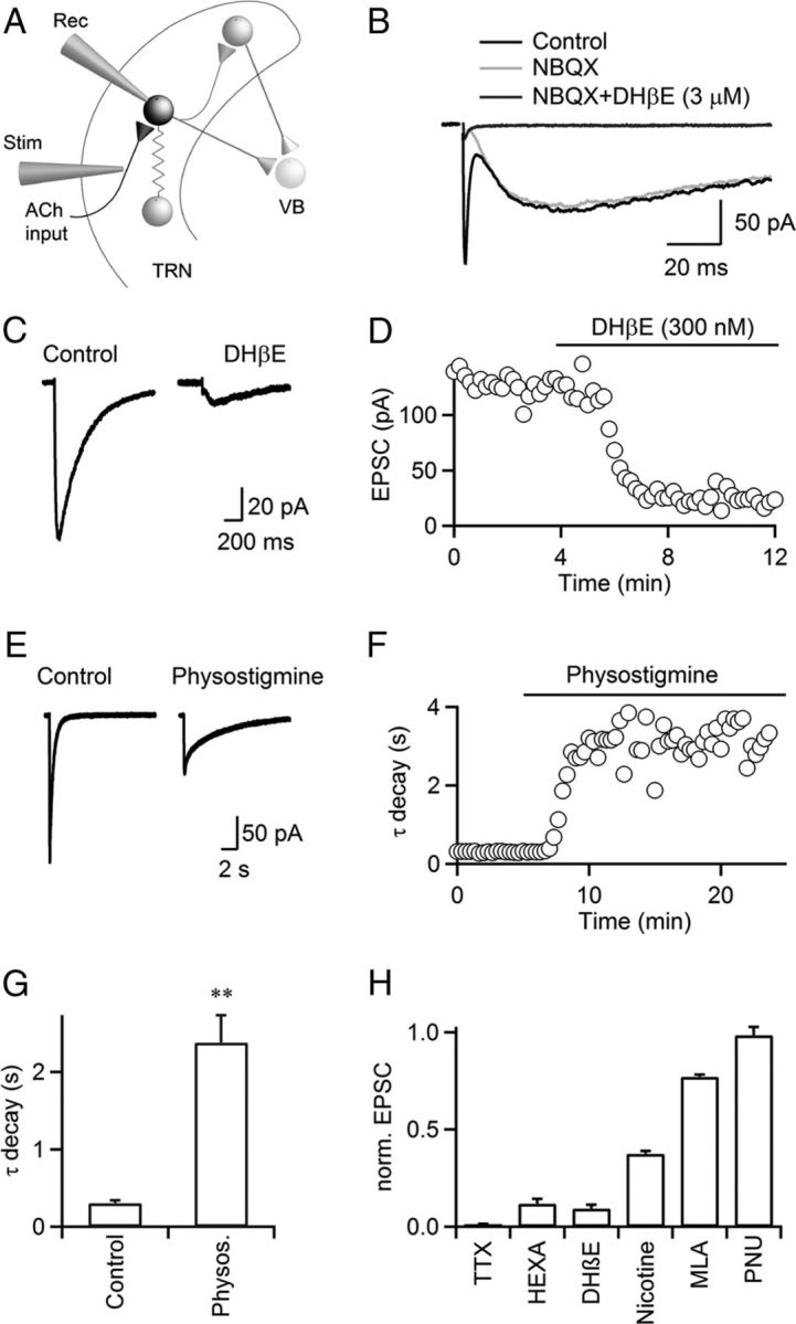
Cholinergic synaptic transmission in the TRN is mediated by α4β2 nAChRs. A, TRN neuronal circuit. TRN neurons were recorded with a Cs-based internal solution. Synaptic inputs were activated with a patch pipette (Stim), placed at lateral distances of 100–200 μm. VB, ventrobasal thalamus; Rec, recording pipette; Stim, stimulus electrode. B, For a representative experiment, single stimuli evoked EPSCs with a fast and a slow component (black trace) in a TRN neuron. The fast component was blocked by the AMPAR antagonist NBQX (10 μm, gray trace), and the slow component was blocked by the nAChR antagonist DHβE (3 μm, dark gray trace). C, D, A representative experiment showing that nEPSCs were blocked by low doses of DHβE (300 nm, C). Time course of nEPSC amplitude during bath application of DHβE (300 nm), for the same cell (D). E, F, A representative experiment showing that application of the AChE inhibitor physostigmine (10 μm) prolonged nEPSC decay and reduced nEPSC amplitude (E). Time course of nEPSC decay during bath application of physostigmine (10 μm, F). Decay was fit by a single exponential function. Experiment was performed in the presence of atropine (10 μm). G, Summary showing that physostigmine (10 μm) increased time constant of nEPSC decay. n = 5, **p < 0.01. Paired Student's t test. H, Summary showing the effect of TTX (500 nm), hexamethonium (HEXA, 100 μm), DHβE (300 nm), nicotine (100 nm), MLA (50 nm), PNU 120596 (PNU, 10 μm) on nEPSC amplitude, normalized to control. n = 5–8.