Figure 5.
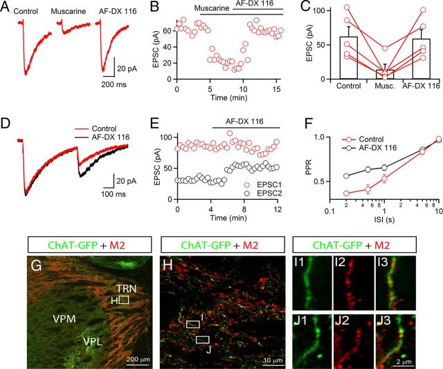
Autoinhibition of ACh release is mediated by presynaptic M2 mAChRs. Recordings were performed with a Cs-based internal solution. A, A representative experiment showing nEPSC suppression by bath application of muscarine (1 μm), reversed by the M2 antagonist AF-DX 116 (10 μm). B, For the same neuron, graph plots time course of nEPSC amplitude before and following application of muscarine and AF-DX 116. C, Summary data showing nEPSC suppression by muscarine (1 μm), reversed by AF-DX 116 (10 μm). n = 5. D, nEPSCs evoked by paired stimuli (500 ms ISI) before (red) and following application of the M2 mAChR antagonist AF-DX 116 (10 μm, black) in a representative recording. E, Time course of EPSC1 (red circles) and EPSC2 (black circles) before and during application of AF-DX 116 (10 μm) for the same neuron as in D. F, Summary showing paired-pulse ratio (EPSC2/EPSC1) for different interstimulus intervals (ISI) in control (red circles) and AF-DX 116 (black circles). n = 5. G, Immunohistochemical staining in sections from ChAT-GFP reporter mice indicates strong expression of M2 mAChRs in the TRN. Expression of ChAT-GFP was labeled by antibodies against GFP (green) and M2 mAChRs were detected by antibodies against M2 mAChRs (red). H, Higher-magnification view for the area indicated in G, showing M2 mAChR is partially overlapping with GFP signal in the TRN. I, J, Examples showing higher-magnification views of the areas indicated in H. M2 mAChR-positive signals (I2, J2, red) colocalize with GFP-positive signal (I1, J1, green), as shown in the overlay in I3 and J3. VPM, ventral posteromedial nucleus of thalamus; VPL, ventral posterolateral nucleus of thalamus.

