Figure 6.
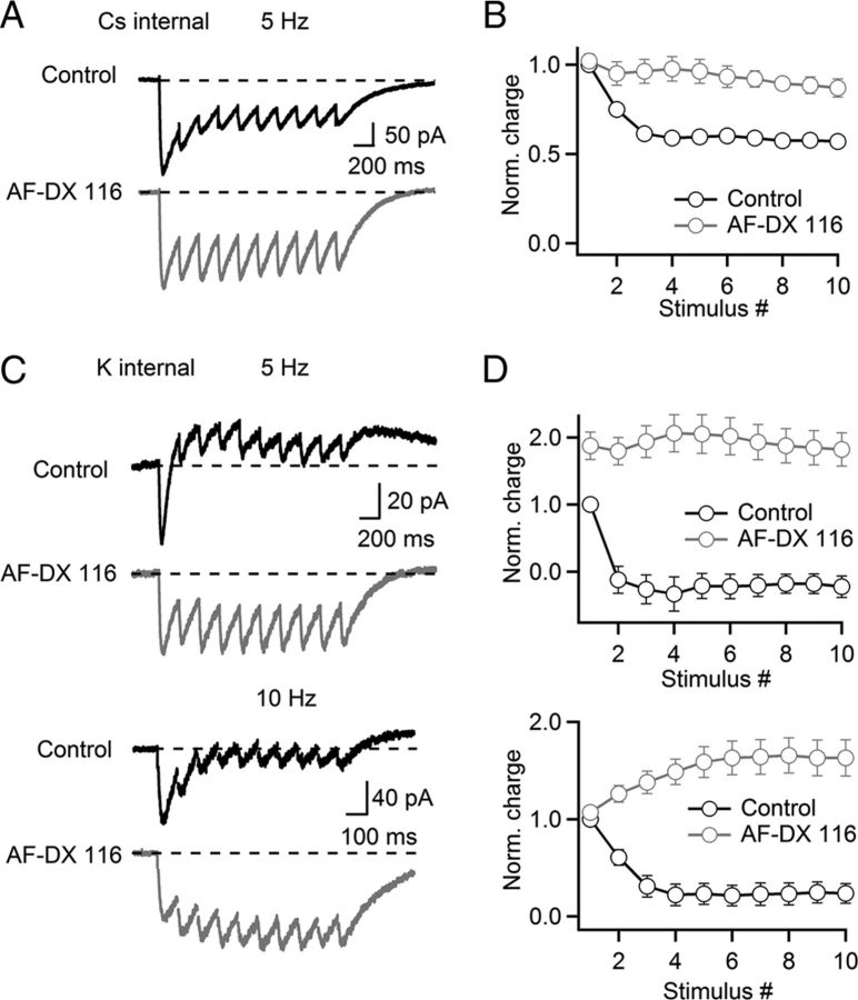
Presynaptic and postsynaptic muscarinic receptors control cholinergic signaling during brief stimulus trains. Recordings in A and B were done with a Cs-based internal solution. For C and D, a K-based internal solution was used. A, A representative experiment showing nEPSCs in response to a train of stimuli (5 Hz, 10 pulses) in control (black trace) and after bath application of AF-DX 116 (10 μm, gray trace). B, Summary data plotting synaptic charge (measured as the area underneath the voltage trace) following each stimulus, normalized to the first EPSC in control (control, black circles; AF-DX 116, gray circles). n = 5 cells. C, Rapid suppression of nAChR-evoked excitation by mAChR activation, blocked by AF-DX 116. Representative experiments showing PSCs in response to trains of stimuli at 5 or 10 Hz in control (black traces) and after bath application of AF-DX 116 (10 μm, gray traces). D, Summary data, plotting net synaptic charge (measured as the area underneath the voltage trace) following each stimulus at 5 or 10 Hz, normalized to the net synaptic charge evoked by the first PSC in control (control, black circles; AF-DX 116, gray circles). n = 8 cells for both frequencies.

