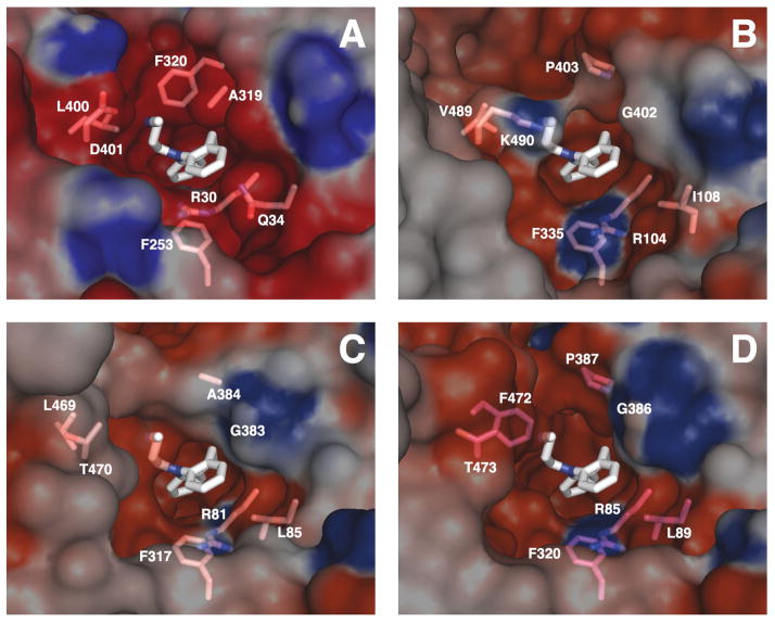
Fig. 3.
Homology models and electrostatic surface potential of desipramine-binding sites in human SERT, NET and DAT. (A). Desipramine-binding site in the LeuT-desipamine crystal structure. Homology model and electrostatic surface potential of desipramine-binding site in (B). hSERT, (C). hNET and (D). hDAT, viewed from within the membrane plane. The equivalent residues of those in LeuT that are in direct contact with desipramine are indicated.

