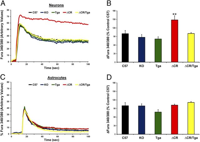
Figure 2.
ΔCR PrP causes abnormal, glutamate-induced Ca2+ influx in NSC-derived neurons. NSCs dissected from mouse embryos at 13.5 d were cultured and propagated as neurospheres, differentiated for 7 d, and then incubated with the Ca2+-sensitive dye fura-2 AM before imaging analysis. Ca2+ influx in response to stimulation with 0.5 mm glutamate was detected by alternating excitation at 340 and 380 nm and monitoring emission at 510 nm. Raw data were collected as the ratio F340/F380, which is directly proportional to the amount of Ca2+ in the cytosol. Neuronal cells (A, B) were discriminated from non-neuronal cells (C, D) on the basis of the expression of the MAP2, which was detected by immunostaining after acquiring Ca2+ influx recordings. Examples of recordings from individual neuronal (A) and non-neuronal (C) cells from the indicated genotypes are shown. B, D, Bar graphs show quantitation of the Ca2+ burst in the different cells, calculated as the difference between F340/F380 before and after glutamate stimulation. Data are reported as percentage of the C57 control. **p < 0.01, one-tailed Student's t test.

