Figure 1.

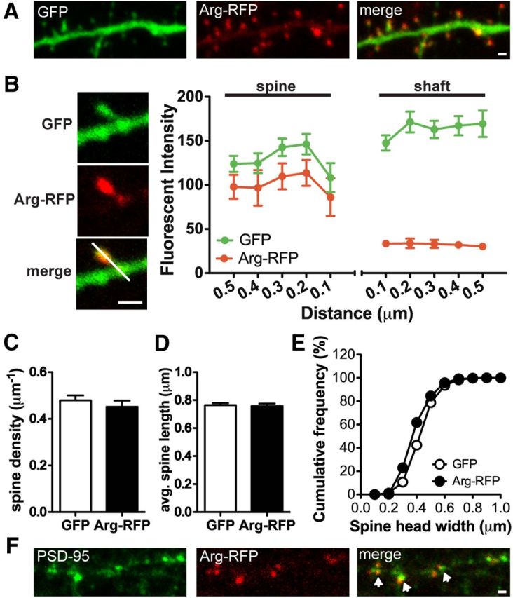
Arg is localized and enriched at dendritic spines. A, Confocal images of a cultured hippocampal neuron at 18 DIV. The neuron was transfected with Arg-RFP and GFP. Scale bar, 1 μm. B, Representative images of a dendritic spine. The quantification of Arg-RFP fluorescence intensity in the dendritic spine versus the dendrite shaft is shown. Fluorescence intensity of Arg-RFP and GFP across the spine and adjacent shaft was measured by a line scan (white line). Note that Arg-RFP is highly enriched in dendritic spines. C, Spine density of GFP- or Arg-RFP-transfected neurons at 15 DIV. D, E, Averaged dendritic protrusion length (D) and cumulative frequency of spine head size (E), of GFP or Arg-RFP-transfected neurons. Note that Arg-RFP does not promote spine formation or changes in the spine length and width. F, Confocal images of an Arg-RFP-transfected neuron immunostained with the postsynaptic marker PSD-95 (green). Note that Arg-RFP (red) is colocalized with PSD-95 puncta (arrows). Scale bar, 1 μm.
