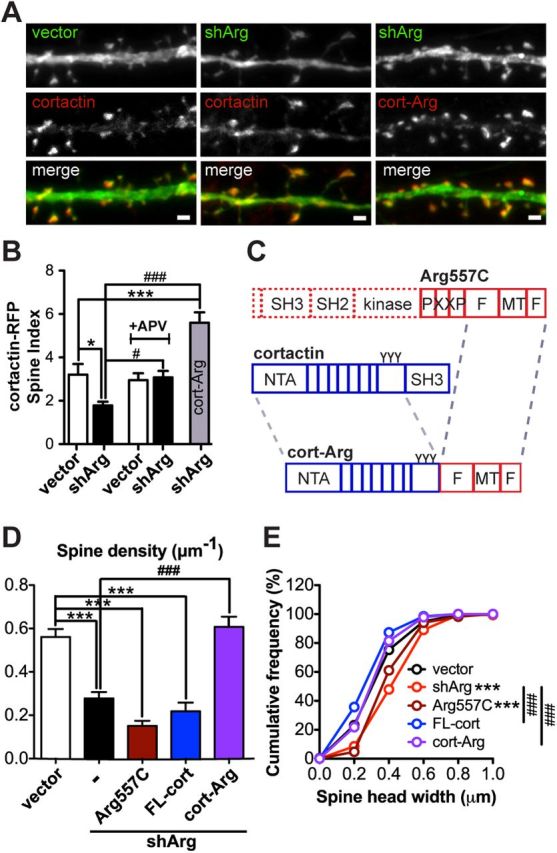
Figure 8.

Forcing cortactin-Arg interactions prevents spine loss. A, Examples of cortactin-RFP (red) or cortactin-Arg-RFP fusion protein (red) localization in control or Arg knockdown neurons. Scale bar, 1 μm. B, Spine enrichment of cortactin-RFP or cortactin-Arg-RFP in neurons. Neurons treated with or without 50 μm APV. n = 16–21 neurons, three independent cultures. *p < 0.05, ***p < 0.001 when compared with control vector; #p < 0.05, ###p < 0.001 when compared with shArg, one-way ANOVA with post-Dunnett's multiple-comparison test. C, Schematic structure of Arg557C, full-length cortactin, and cortactin-Arg (cort-Arg) fusion protein. The cort-Arg fusion protein is constructed by fusing cortactin lacking its SH3 domain with Arg lacking the N-terminal and PXXP motif (Arg-668C). D, Spine density of vector- or shArg-transfected neurons. Arg knockdown neurons were complemented with the Arg557C domain (dark red), full-length cortactin (FL-cort; blue), and cort-Arg fusion (purple). Cort-Arg restores spine density in Arg knockdown neurons. ***p < 0.001 when compared with control neurons; ###p < 0.001 when compared with Arg knockdown neurons, one-way ANOVA with post-Dunnett's multiple-comparison test. n = 28–42 neurons, four independent cultures. E, Cumulative frequency of spine head width of vector- or shArg-transfected neurons complemented with constructs as indicated in D. ***p < 0.001 when compared with control; ###p < 0.001 when compared with shArg, Kolmogorov–Smirnov test.
