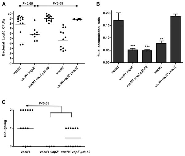
Figure 6. VopZ Makes Distinct and Genetically Separable Contributions to V. parahaemolyticus Colonization and Pathogenesis within the Infant Rabbit Small Intestine.
Bacterial colonization (cfu/g) in intestinal tissue (A) and intestinal fluid accumulation ratios (B) for infant rabbits infected with the indicated strains of V. parahaemolyticus at ~38 hr postinfection. Lines in (A) show geometric means. Means and SEM, based on at least nine rabbits, are shown in (B). Data for the vscN1 and vscN2 strains were previously published (Ritchie et al., 2012). Tissue sloughing (C) in tissue from distal intestines of infected infant rabbits was scored as described (Ritchie et al., 2012); median values are indicated. Statistical significance was assessed with one-way ANOVA and Bonferroni’s posttests: *p < 0.05, **p < 0.01, ***p < 0.001, or as indicated on the graphs.

