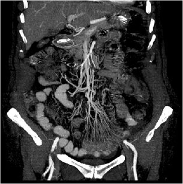
Figure 2.
Acute arterial mesenteric ischemia. Contrast-enhanced MDCT 2D reconstruction on coronal plane in early phase: the CT shows the presence of emboli or thrombi as filling defect in the lumen of the artery. If they are small and peripherally localized, the identification can be difficult. The loops of injured small bowel are contracted in consequence of spastic reflex ileus and intestinal wall shows lacking of/poor enhancement. The mesentery is bloodless, due to reduction in caliber of the vessels and apparently in number

