Figure 4.
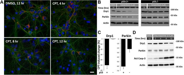
CPT suppresses Drp1 and parkin protein expression in postnatal cortical neurons in a p53-dependent manner. A, p53+/+ neurons were treated with DMSO (control, 12 h only) or CPT (5 μm) 3 d after plating for 4, 8, and 12 h, fixed and immunostained for Drp1(red) and Tuj1 (green) with Hoechst 33258 used to delineate nuclei (blue). Scale bar, 10 μm. The images are representative of four independent experiments. B, p53+/+ or p53−/− neurons were treated with DMSO (control, 12 h only) or CPT (5 μm) 3 d after plating and 2, 4, 8, and 12 h later protein samples were prepared and analyzed for expression of Drp1 and parkin by Western blot. β-Actin was used as an internal loading control. C, Western blot results including those shown in B were quantified. In p53+/+ neurons the average CPT-induced decline in Drp1 and parkin expression at 12 h relative to DMSO control (and normalized to β-actin) was 50.53 ± 6.60% (mean ± SEM, p < 0.001, Student's t test, N = 20) and 60.74 ± 5.32% (p < 0.001, N = 9), respectively. CPT did not reduce Drp1 levels in p53−/− neurons (0.53 ± 0.53%, N = 3), but did induce a significant reduction in parkin expression (22.3 ± 7.02%, p < 0.05, N = 2), although it was substantially less than that observed in CPT-treated p53+/+ neurons. D, p53+/+ neurons were treated with DMSO (control, 12 h only) or staurosporine (0.5 μm) 3 d after plating and 2, 4, 8, and 12 h later protein samples were prepared and analyzed for expression of Drp1, parkin, and activated caspase-3 by Western blot. β-Actin was used as an internal loading control. Blots are representative of at least three separate experiments.

