Figure 6.
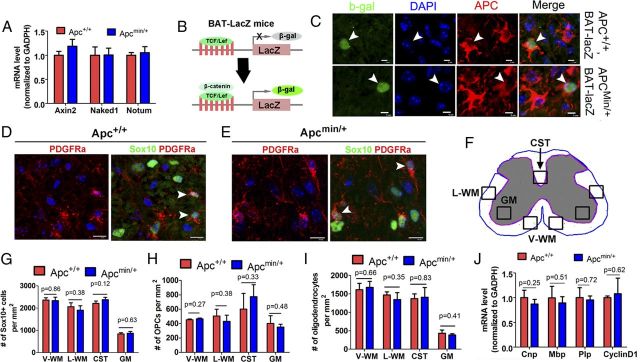
Wnt signaling and oligodendrocyte differentiation are not perturbed in Apcmin/+ mice. A, MRNA levels of Wnt target genes Axin2, Naked1, and Notum in Apcmin/+ (n = 4) and Apc+/+ WT (n = 4) spinal cord at P7. B, Schematic drawing showing the mechanism of β-gal expression in BAT-lacZ Wnt reporter mice. C, β-Gal expression in APC+ cells from BAT-lacZ/Apc+/+ (top) and BAT-lacZ/Apcmin/+ mice (bottom) at P7. D–E, Representative confocal images showing Sox10+ oligodendroglial lineage cells and Sox10+/PDGFRα+ OPCs in the WM of Apc+/+ and Apcmin/+ spinal cord at P7. F, Schematic image depicting the sampling locations for G–I. V-WM, ventral WM; L-WM, lateral WM; CST, corticospinal tract. G–I, Quantifications of the frequency of Sox10+ panoligodendroglial lineage cells (G), Sox10+/PDGFRα+ OPCs (H), and Sox10+/CC1+ oligodendrocytes (I) in the spinal cord at P7 (n = 4 in each group). J, MRNA expression level of Cnp, Mbp, Plp, and cyclin D1 by qRT-PCR in P7 Apc+/+ and Apcmin/+ spinal cord (n = 4 in each group). Scale bars: C, 5 μm; D, E, 10 μm.

