Fig. 3.
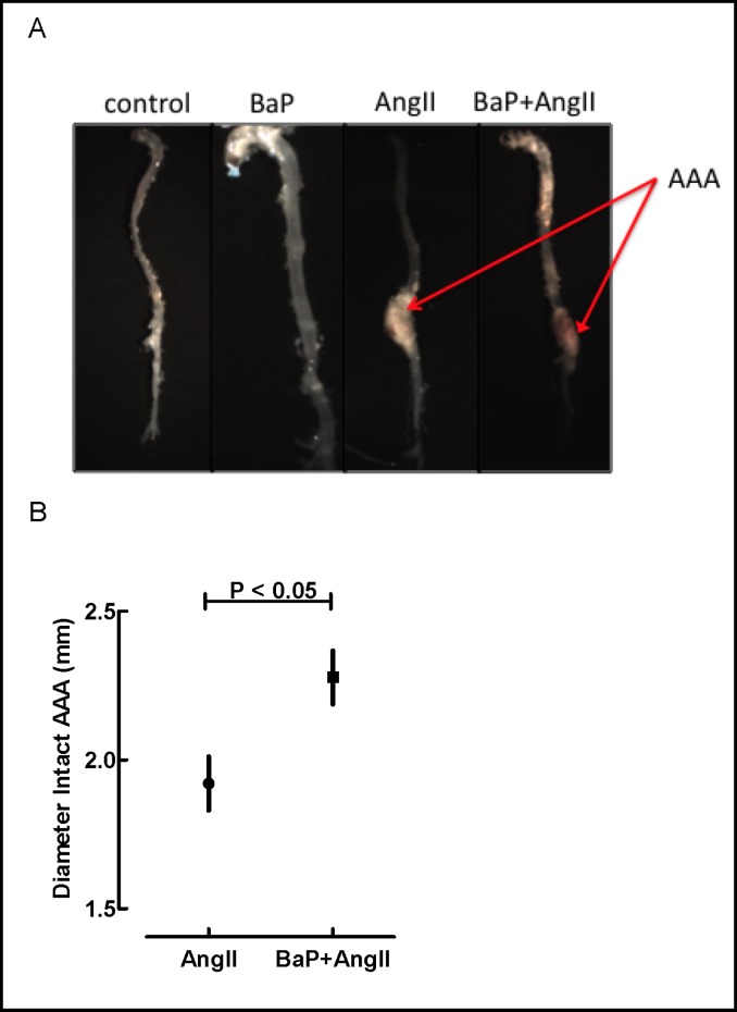
Anatomical evaluation of the aorta. A) Representative gross images of aortas from control, BaP, AngII and BaP+AngII groups. AAA is indicated (arrow) and increased atherosclerosis is noted in the arch of aortas from BaP and BaP+AngII groups. Images are derived from the weekly-dosing experiment. B) Sizes of AAA. In the daily-exposure arm, the maximum diameter of intact AAA was measured using a digital caliper under microscope guidance. Diameters of AAA in BaP+AngII treated mice were significantly larger (2.3 mm ± 0.1, N=8) than diameters of those in AngII treated mice (1.9 mm ± 0.1, N=10) (p < 0.05). We did not observe any significant differences in the diameter of intact AAA in the weekly-dosing experiment (data no shown).

