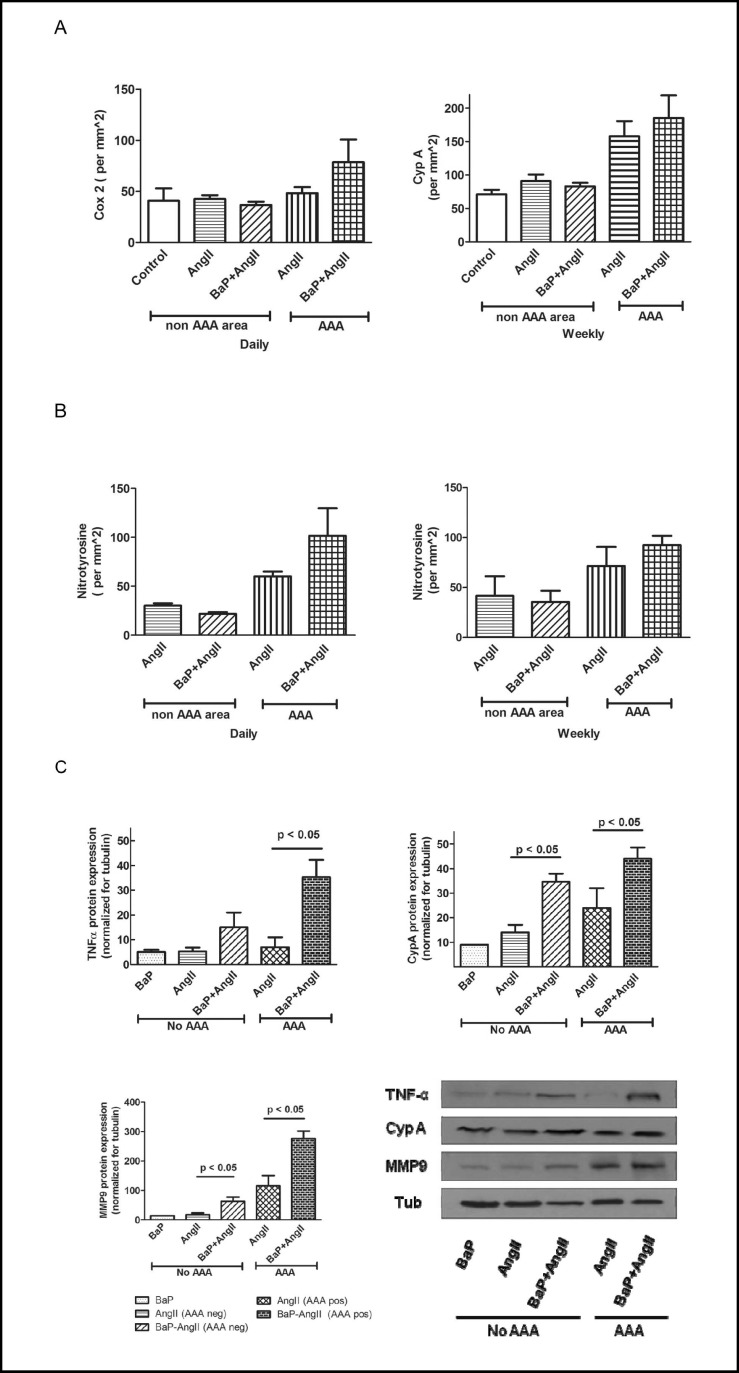
Fig. 5.
Effects on markers of inflammation, proteolysis, and oxidant stress. A) Cox-2 positive cells are present in the aortic wall with a trend toward increased positive cells in AAA sites of BaP+AngII (n=4) versus AngII (n=5) treated mice. Also, increased levels of CypA which promotes inflammation and is expressed by vascular smooth muscle cells were observed at AAA sites of mice in both AngII (n=5) and BaP+AngII (n=4). There was no statistically significant difference in the expression of Cox2 and CypA in both groups. Similar trends were noted for CypA in the daily exposure experiment (data not shown). B) Increased oxidant stress is suggested (p= NS) by the nitrotyrosine stain of AAA sites in the daily (AngII n= 5, BaP+AngII n= 3) but not the weekly (AngII n= 4, BaP+AngII n= 4) exposure experiments. Cross sections used for AAA and non AAA were from the same aortas. C) The expression of TNF-α, CypA, and MMP9 protein were significantly higher in aorta from mice in BaP+AngII compared to Ang II group independent of the presence of AAA (p < 0.05). Results shown are data from 2 sets of groups, each group representing a triplicate assessment of protein pooled from 3 aortas from AAA negative and AAA positive mice, respectively. Data shown are only for daily exposure experiment.

