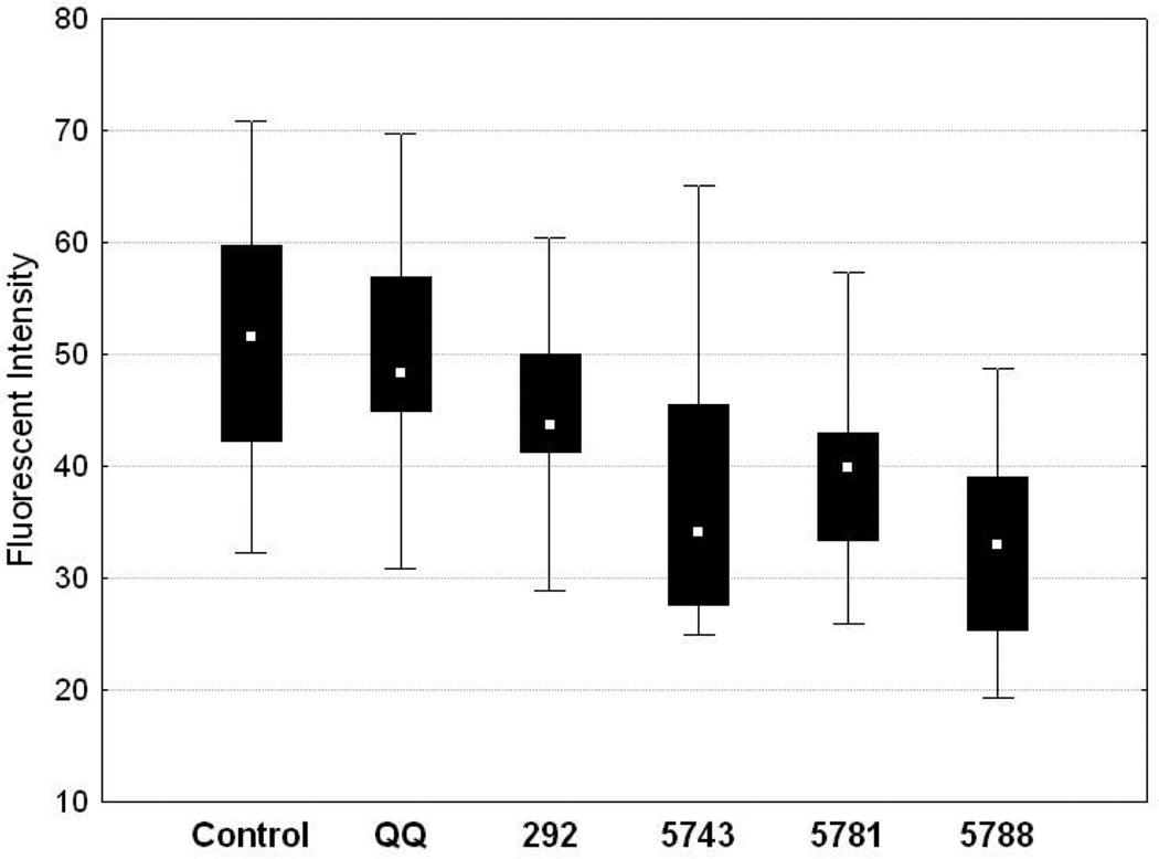
Figure 2.
Treatment of HeLa cells with PBD inhibitors leads to reduced localization of PLK1 to the centrosomes. The Y-axis represents arbitrary fluorescent intensity units. Control represents fluorescence intensity data from untreated cells. QQ represents the data from cells treated with transfection reagent alone. The remainder represent data from cells transfected with the following peptides: 292 (LLCS[pT]PNGL), 5743 (Ac-PLHS[pT]A), 5781 (Ac-PLHS[pT]A), 5788 (3G1-S[pT]PNGL). Statistically significant decreases in fluorescent intensity were observed for 5743, 5781 and 5788.

