
Figure 2.
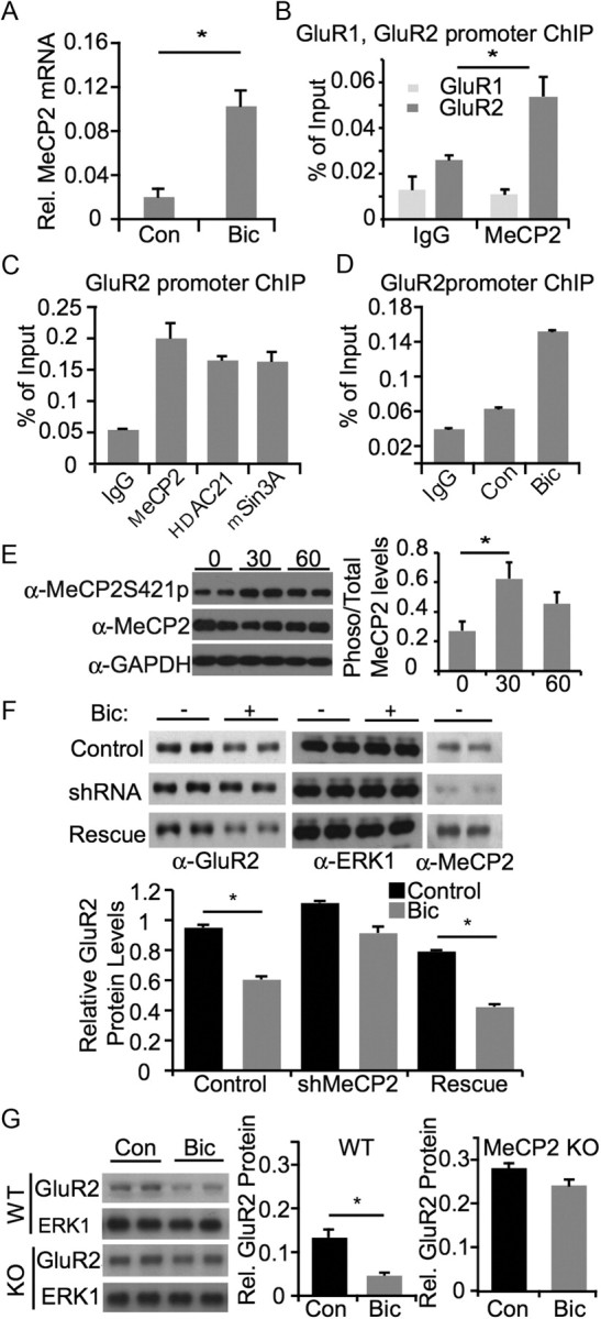
MeCP2 is required for activity-dependent downregulation of GluR2 expression. A, Assessment of MeCP2 mRNA after bicuculline stimulation. Rat E18 cortical neurons were cultured for 16 DIV and stimulated with bicuculline for 6 h. Neurons were lysed for collecting total RNA and reverse-transcribed for real-time PCR analysis with MeCP2-specific primers. B, MeCP2 chromatin immunoprecipitation from E18 + 7 DIV rat cortical cultures. After immunoprecipitation with anti-MeCP2 antibody, PCRs with endogenous GluR1 and GluR2 promoter primers were used to amplify promoter-specific segments. Real-time quantitative PCR was performed and signals were normalized as percentage of input to determine promoter occupancy. C, MeCP2, HDAC1 and mSin3A chromatin immunoprecipitation from rat cortical neurons under basal conditions. After immunoprecipitation with anti-MeCP2, anti-HDAC1 and anti-mSin3a antibody, PCRs with endogenous GluR2 promoter primers were used to amplify GluR2 promoter-specific segments. Real-time quantitative PCR was performed and signals were normalized as percentage of input to determine promoter occupancy. D, Relative binding of MeCP2 to the GluR2 promoter in unstimulated and bicuculline-stimulated neurons examined using chromatin immunoprecipitation. E, Left, Western blot from hippocampal cultures after exposure to bicuculline for 0, 30, or 60 min. Phosphorylated MeCP2 is detected using a Serine 412 phospho-specific antibody. Right, quantification of ratio of total MeCP2 to MeCP2 phosphorylated at Serine 421 at 0, 30, or 60 min bicuculline exposure. Asterisk signifies p < 0.05 by ANOVA. F, Assessment of changes in GluR2 expression following bicuculline stimulation, with and without MeCP2. Rat P0/P1 hippocampal neurons were cultured for 8 DIV and infected with lentivirus containing control shRNA, MeCP2 shRNA, or MeCP2 shRNA with an shRNA-resistant MeCP2 construct (rescue). Cultures were stimulated with bicuculline for 48 h where indicated, and lysed for Western blot analysis with specified antibodies. Asterisk indicates p < 0.05 using t test. G, Assessment of changes in GluR2 protein levels following bicuculline stimulation in wild-type and MeCP2 knock-out neurons. P7 hippocampal slices were prepared from wild-type and MeCP2 knock-out mouse and cultured for 3 d. After stimulation with bicuculline for 48 h, neurons were lysed for Western blot analysis with antibodies as indicated.