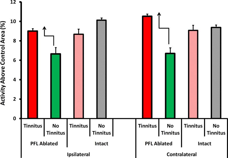
Figure 8.
Summary of the MEMRI imaging results of Experiment 2 animals depicted in Fig. 7. The only consistent activity pattern differences, tinnitus vs. no-tinnitus, that appeared between intact and ablated animals, was evident in the MGB. Ablated rats that developed tinnitus had elevated MGB activity with respect to ablated rats that did not develop tinnitus (dark red vs. dark green bars). In contrast, intact rats that developed tinnitus had the same level of MGB activity as intact rats without tinnitus (light red vs gray bars). The activity pattern of other auditory areas was similar across all four groups (not shown, see text). Error bars shown the mean deviation.

