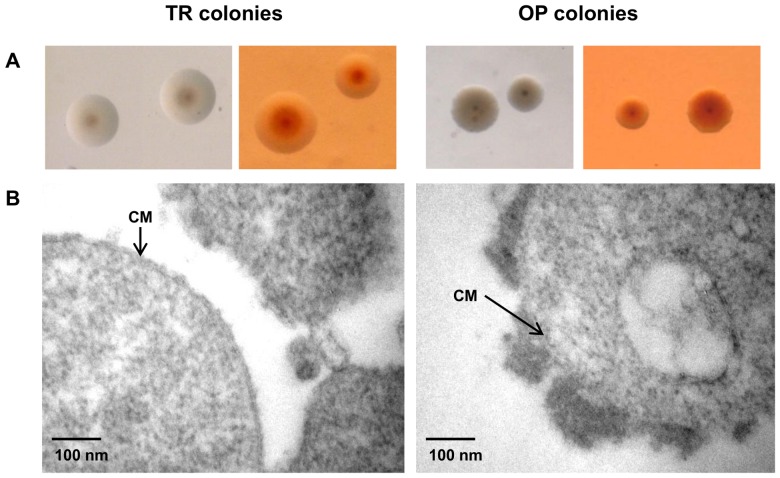
Figure 4. PG1T colony opacity is related to the presence of capsular material.
A: Colony morphology of PG1T opaque (OP) and translucent (TR) colony variants grown on PPLO supplemented agar with (right) or without (left) Congo red. Magnification: x30. B: Electron microscopy of Ruthenium red-stained PG1T OP and TR colonies. CM: cytoplasmic membrane.

