Figure 2.
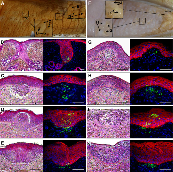
Distribution and structure of lingual sensory organs in crocodylians. Macrophotographs of the C. niloticus(A) and Caiman crocodilus(F) tongue showing the presence of different types of organs over its surface: salt glands (B), large (C and H) and small (G) ISO-related structures, taste buds (D and I), and large epithelial thickenings (E and J). Parasagittal sections were stained with H&E (left panels) or processed for double immunostaining (right panels) of pan-cadherin (general epithelial marker, red) and acetylated tubulin (neuronal marker, green). Cell nuclei were counterstained with 4',6-diamidino-2-phenylindole (blue). Magnification bars: 100 μm.

