Figure 5.
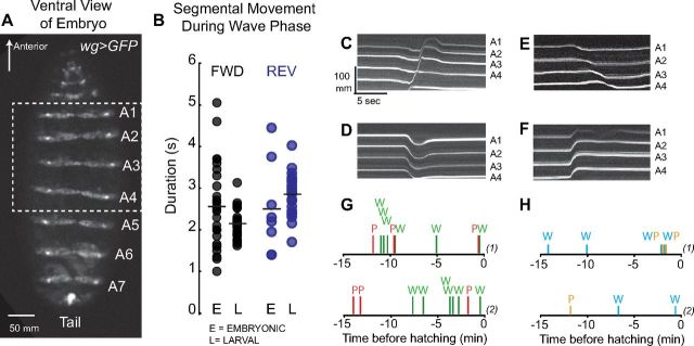
Embryonic wave phase and embryonic visceral piston phases can occur independently in embryos. A, We used wg>GFP to monitor segmental movements in embryonic stages. A representative image of a wg>GFP embryo at stage 17e/f, after the trachea have been inflated is shown. Body wall segments A1–A7 are labeled; the dashed box shows the region of the embryo viewed in C–F. View is ventral with anterior to the top. B, We measured the time it took for a single segment to move in the direction of the wave phase during embryonic and larval wave phases. Duration of movement during forward embryonic wave phase (black dots in E column), during forward larval wave phase (black dots in L column), during reverse embryonic wave phase (blue dots in E column), and during reverse larval wave phase (blue dots in L column) are plotted. Means are indicated with black lines. Note for both forward and reverse wave phases the ranges overlap, with the embryonic ranges more wide-spread. C–F, Kymographs show embryonic wave phases (C, E) and embryonic visceral piston phases (D, F). During an embryonic forward wave phase (C) each segment moves toward the head. During an embryonic reverse wave phase (E) each segment moves toward the tail. There are two embryonic visceral piston phases: in one type (F) all segments moved in unison toward the head, which is likely to correspond to a larval reverse visceral piston phase because the center of mass cannot translocate across the substrate when the embryo is encased in the egg. In the other type (D), all segments moved in unison toward the tail, which is likely to be the embryonic equivalent of a larval forward visceral piston phase. We noted that each of these phases could occur independently, without any other segmental movement within 5 s. G, H, We scored the sequence of tail to head embryonic waves (green bars, W), and head to tail embryonic piston phases (red bars, P), which likely correspond segmental movements seen during a forward larval crawl. We also scored the sequence of head to tail embryonic waves (blue bars, W), and tail to head embryonic piston phases (orange bars, P), which likely correspond to segmental movements seen during a reverse larval crawl. Examples from two embryos [denoted as (1) and (2)] are shown. Note that each movement can occur independently.

