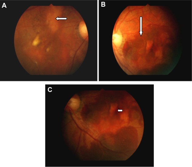
Figure 2.

Fundus findings in patient 8 with idiopathic polypoidal choroidal vasculopathy. Right eye (A) with resolved subretinal blood and macular degeneration. (A) subretinal orange lesions (medium arrow); The left eye (B) shows fresh subretinal hemorrhage (long arrow); and (C) retinal pigment epithelial detachment (short arrow). Note the absence of drusen seen in age-related macular degeneration.
