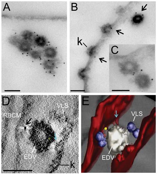
Figure 8. Immuno-EM of EDVs with associated PfEMP1.
EqtII-permeabilized 3D7 strain parasites (A-C) and PfEMP1B-GFP transfectants (ITK strain) (D, E) were labeled with antibodies recognizing the ATS (A-C) or GFP (D, E) and prepared for EM (A-C) or tomography (D, E). Arrows indicate gold particles. The RBC membrane is rendered in red, VLS in blue, EDV in white and a gold particle in yellow. A region that may represent an RBC cytoskeleton extension is indicated with aqua arrows. Scale bar = 100 nm.

