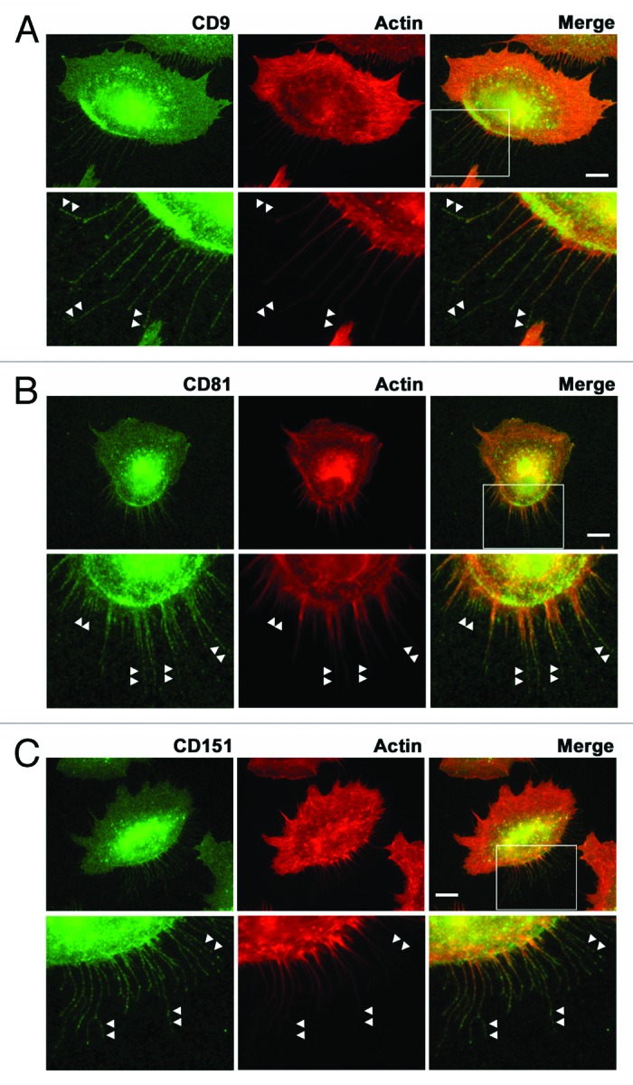
Figure 3. Localization of tetraspanins in retraction fibers in migrating cells. A549 cells migrating on laminin-511 were stained with anti-CD9 (A), anti-CD81 (B), and anti-CD151 (C) antibodies (green) and rhodamine-phalloidin (red) as described in Materials and Methods. The lower panels show high magnification views of the boxed areas. Arrowheads indicate tetraspanin-positive but F-actin-negative regions in retraction fibers. Bars represent 10 μm.
