
Figure 2. The α4 nAChR is colocalized with TH-positive dopaminergic neurons of the VTA.
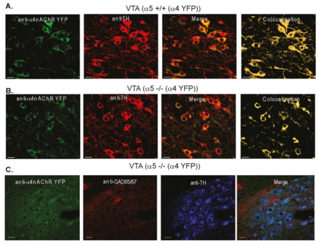
Representative immunofluorescence images from α5+/+(α4 YFP) (A) and α5-/-(α4 YFP) (B and C); VTA showing α4 nAChR-YFP expression (green), tyrosine hydroxylase (TH) (red) expression, the merged images (green + red) and the colocalization (yellow); VTA showing α4 nAChR-YFP expression (green), GAD65/67 (red), tyrosine hydroxylase (TH) (blue) expression, and the merged images (green + red + blue). Scale bar is 30µm.