
Figure 3. The α5 subunit controls the strength of nicotinic currents mediated by the α4*-containing nAChRs in VTA dopaminergic neurons.
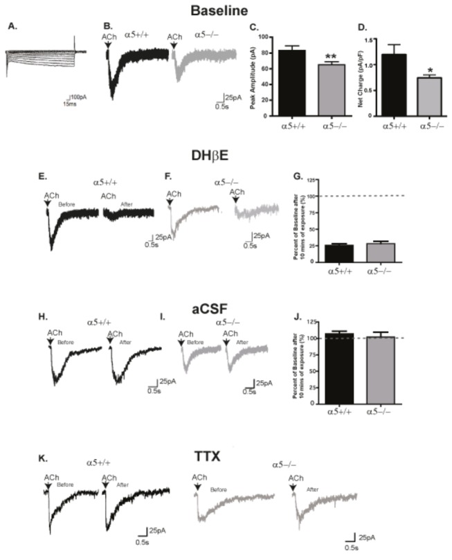
(A) A typical Ih current. (B) Sample voltage clamp traces of peak inward current of DA neurons to a 300 ms ACh (1mM) puff in α5+/+ (black) and α5-/- mice (gray). (C) The average ACh-induced peak current amplitude was reduced in dopaminergic neurons from α5-/- mice in comparison to α5+/+ mice. (D) The average net charge was reduced in dopaminergic neurons from α5-/- mice in comparison to α5+/+ mice. (E and F) Both α5+/+ (black) (E) and α5-/- (gray) (F) mice showed a nearly complete reduction in the nicotinic currents after 10 min of α4 nAChR antagonist DHβE (2µM) treatment indicating that the responses are mediated by the α4* nAChRs. (G) The percent reduction from baseline following DHβE treatment were similar for α5+/+ and α5-/- mice. (G and H) Currents were stable to 300ms ACh puffing every 2 min for 20 min in neurons exposed to aCSF in both α5+/+ (black) (H) and α5-/- (gray) (I). (J) There was no significant percent reduction from baseline in both genotypes. (K) TTX (2 µM) had no effect on the current in both α5+/+ and α5-/- mice. In C & D, n = 57-61 cells across 45-50 animals, F, n =7 cells across 6 animals and in I, n=6 cells across 5-6 animals. The values in C are mean peak amplitude ±SEM (two-tailed unpaired t-test, **p<0.01). The values in F&I are reported as mean percent of baseline ±SEM (two-tailed unpaired t-test). The calibrations for the current trace are 100pA, 15 sec (A) and 25pA, 0.5sec (B, E and H).