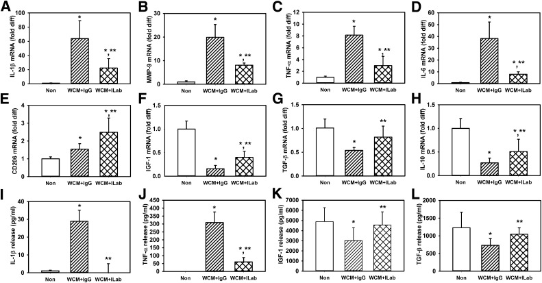
FIG. 3.
IL-1β–neutralizing antibody downregulates diabetic wound–conditioned medium–induced proinflammatory phenotype and upregulates healing-associated phenotype in cultured macrophages. Bone marrow–derived macrophages from wild-type mice left nonstimulated (Non) or stimulated with day 10 db/db wound-conditioned medium (WCM) along with control IgG or stimulated with IL-1β–neutralizing antibody (ILab); expression of proinflammatory markers (A–D) IL-1β, MMP-9, TNF-α, and IL-6 and healing-associated/anti-inflammatory markers (E–H) CD206, IGF-1, TGF-β, and IL-10 were measured by real-time PCR. In addition, release of (I–L) IL-1β, TNF-α, IGF-1, and TGF-β was measured using ELISA. For all graphs, bars = mean ± SD. For these experiments, a separate set of bone marrow–derived macrophages (each harvested from a different mouse) was used for each of two experiments and wound-conditioned medium was generated from three mice per experiment, with n = 6 for each condition. *Mean value significantly different from that for nonstimulated controls. **Mean value significantly different from that for conditioned medium plus IgG-treated samples, P < 0.05. diff, difference.

