
FIG. 8.
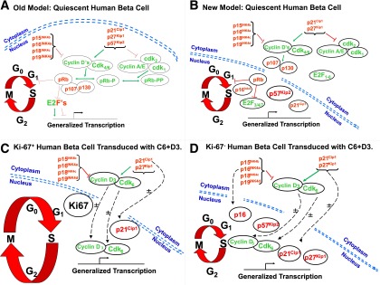
Previous and revised models of the G1/S cell cycle transition regulation in human β-cells. The red arrows indicate inhibition, the green arrows indicate activation, and the dotted black lines indicate trafficking. A: A previous model of G1/S control in human β-cells (2–13) in which most cell cycle molecules were assumed to be present in the nucleus. B: A revised model for G1/S molecule distribution in quiescent human β-cells: most G1/S molecules are cytoplasmic, and the only other frequently nuclear molecules are the cell cycle inhibitors, pRb, p57, accompanied in occasional β-cells by p16, or, in some preparations, p21. C: G1/S molecules in Ki67+ human β-cells that recently have proliferated or are proliferating. The larger G1/S circle, as compared with the others, indicates that these cells have entered the cell cycle. After transduction, cdk6 and cyclin D3 transit to the nuclear compartment in association with p21. D: G1/S molecules in human β-cells that remain Ki67− (fail to proliferate) despite transduction with cdk6 and cyclin D3. The cdk6 and cyclin D3 still enter the nucleus in many β-cells but are accompanied variably by p16, p21, p27, or p57, all or some of which may attenuate the induction of proliferation. The ± symbols in C and D indicate that these movements may or may not occur in a given β-cell after cdk6–cyclin D3 transduction.