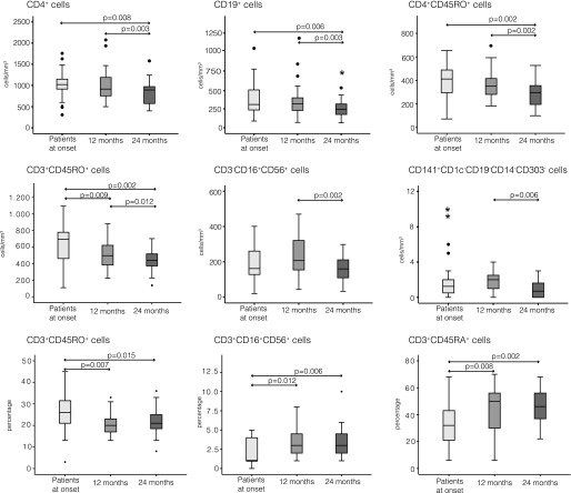
FIG. 2.
Immunological follow-up in type 1 diabetes. Children affected by type 1 diabetes were studied at diagnosis and at 12 and 24 months after disease onset. Box plots show the distribution of CD4+ T cells, CD19+ B cells, CD4+CD45RO+ T cells, CD3+CD45RO+ T cells, CD3-CD16+CD56+ cells, and CD141+ cells and percentages of CD3+CD45RO+ T cells, CD3+CD16+CD56+ cells, and CD3+CD45RA+ T cells in patients at onset and after 12 and 24 months. Data are shown as median (horizontal line in the box) and Q1 and Q3 (borders of the box). Whiskers represent the lowest and the highest values that are not outliers (i.e., data points below Q1 − 1.5 × IQR or above Q3 + 1.5 × IQR) or extreme values (i.e., data points below Q1 − 3 × IQR or above Q3 + 3 × IQR). Dots represent outlier values and asterisks represent extreme values. Q1 = 25th percentile; Q3 = 75th percentile; IQR (interquartile range) = Q3–Q1.

