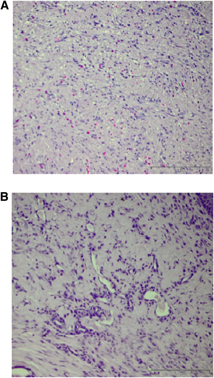
FIG. 5.
Representative images of neovasculature in control wounds (A) and wounds treated with 1,000,000 MSCs seeded in a collagen scaffold (B). Tissue samples are fixed in paraffin at 5-µm depth and stained with hematoxylin and eosin. Scale bar, 200 µm. An increased blood vessel density and reduced radial diffusion distance are evident in the wounds treated with MSCs and collagen as compared with untreated wounds, as evident in B and A, respectively. (A high-quality color representation of this figure is available in the online issue.)

