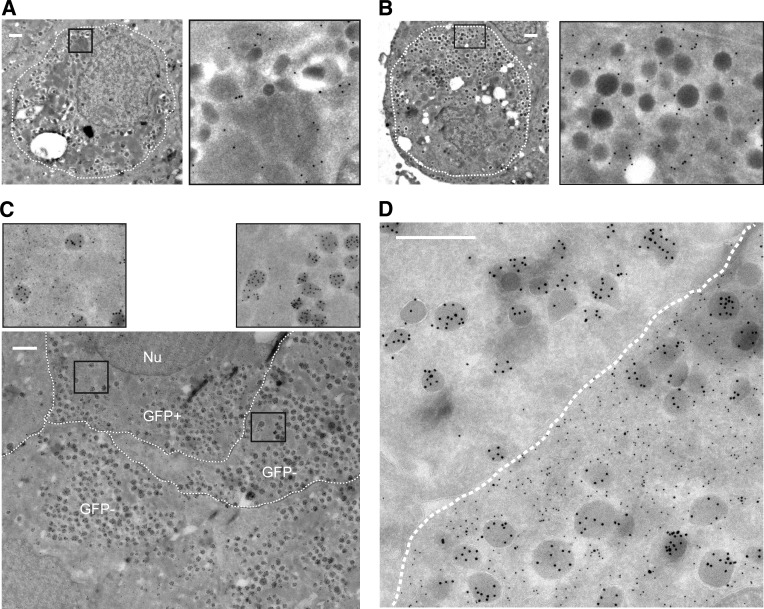
FIG. 4.
Converted cells are regular α-cells based on ultrastructural morphology. A and B: Immunogold labeling for GFP (15-nm gold particles) is present as black dots in both cells with granules with a typical crystalline structure containing insulin (A) and cells with more homogenous dark and dense noninsulin granules (B). C and D: Double immunogold labeling for GFP (10-nm gold particles) and glucagon (15-nm gold particles) shows the presence of α-cell granules in both GFP+ (converted) and GFP− cells (C). Immunogold labeling for GFP is absent within granules (D). The borders between the cells are marked manually by a broken line (Nu = nucleus). All scale bars = 500 nm.

