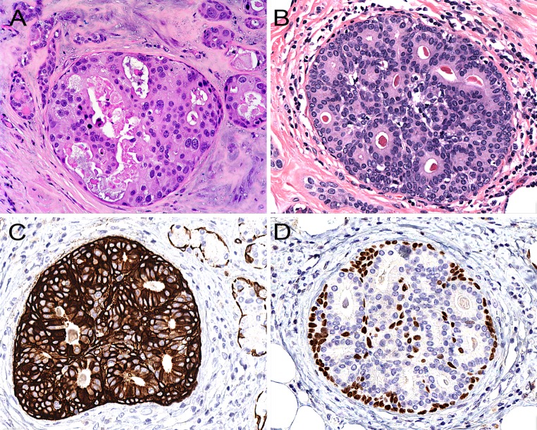
Fig. 7.
a When intraductal proliferations in sclerosing polycystic adenosis display obvious nuclear pleomorphism and luminal necrosis, the diagnosis of ductal carcinoma in situ is easy. b A “low-grade” cribriform intraductal proliferation with distinct luminal differentiation similar to mammary atypical ductal hyperplasia/low-grade ductal carcinoma in situ. c The lesional cells are frequently strongly positive for cytokeratin 5/6, and some cells may also be positive for p63 (d), a feature that differs from ADH/low-grade DCIS in the mammary gland

